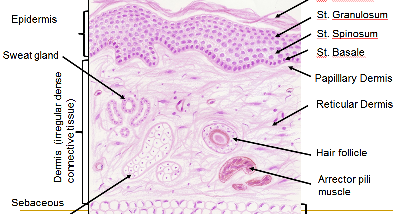
Thin Skin Histology Diagram
Histology drawings: skin (integumentary system) Skin – normal histology – nus pathweb :: nus pathweb Skin (integumentary system)
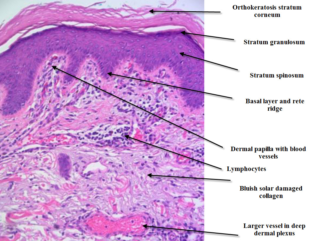
Histology (Skin) - Part 1
Siteground system page not active Hematoxylin histology integumentary epidermis eosin trichrome alive Histology slides database: january 2014
Skin anatomy and wound healing
Diagram of thin skin structureHistology (skin) Histology hypodermis guide thin leeds sweat glands pili arrector gland sebaceous microscopeSkin histopathology simple made introduction dermatopathology neoplastic dermpath dr.
Histology integumentary 收藏Histology (skin) Histology thin objective microscopySkin histology anatomy hair scalp dermis follicle human diagram follicles wound healing microscope slide tissue glands layers epidermis integumentary thick.

Histology dermis tissue epithelial sebaceous glands physiology corpuscles krause zapisano
Skin reading.php labHistology skin thin system integumentary drawings human anatomy thick section cross mallory trichrome slides cutis 40x nervous renal between body Histology skin thin diagram slides resolution highScalp (skin).
Skin epidermis layers histology labHistology of skin Skin histology anatomy hair scalp dermis follicle human diagram follicles microscope slide tissue layers wound healing glands thick microscopic structuresHistology drawings: january 2014.

Histology dermis tissue epithelial physiology sebaceous appendages
Histology integumentary anatomy stain tissue types layers mallory trichrome cutisDermpath made simple Histology nus pathweb annotations.
.


